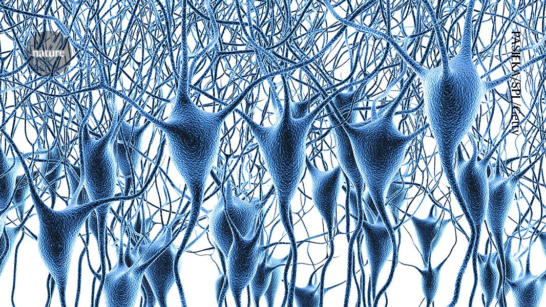
A detailed brain map describes gene activity in neurons (artist’s illustration) as well as immune and vascular cells in the prefrontal cortex.Credit: PASIEKA/SPL/GettyResearchers have produced the largest map to date of gene activity in the human prefrontal cortex1, a brain area that supports planning, decision-making and behavioural and emotional regulation. The landmark map draws on sequencing data from more than six million individual cells from almost 1,500 people to inform the study of neurodegenerative and psychiatric diseases in unprecedented detail.The research was published today in a collection of eight studies, including three papers in Nature1,2,3.“The scale of this project and the amount of work required to assemble it are genuinely impressive,” says Zhichao Miao, a computational biologist at the Guangzhou National Laboratory in China who was not involved in the work. “Single-cell studies of the human brain have traditionally been limited to relatively small numbers of individuals. Pushing us into a population-scale setting changes the kinds of questions we can ask.”Brain planning centrePanos Roussos, the director of the Center for Disease Neurogenomics at the Icahn School of Medicine at Mount Sinai in New York City and a co-author on all eight papers, says that the work is the culmination of a massive effort that first began in 2019. The PsychAD Consortium, an NIH-funded partnership between several US institutions, was established with the goal of connecting genetic variation, ageing and disease to changes in specific brain cells.The group focused on the prefrontal cortex, Roussos says, because of its crucial role in working memory and ‘executive function’ — high-level processes that direct planning, focusing and multitasking. Disruptions to a specific subregion called the dorsolateral prefrontal cortex, Roussos adds, are implicated in several psychiatric disorders and types of dementia.Biggest brain map ever details huge number of neurons and their activityTo build their maps, the team studied tissue from nearly 1,500 people whose brains were donated to science after their deaths. The donors ranged from infants to an individual aged 108, had diverse ancestries and included both neurotypical controls and people diagnosed with one of eight brain disorders: Alzheimer’s disease; dementia with Lewy bodies; Parkinson’s disease; vascular dementia; tauopathy; frontotemporal dementia; schizophrenia; or bipolar disorder.The researchers performed a type of analysis called single-cell RNA sequencing, which details the RNA transcripts active in an individual cell at the time it is sampled. This gives scientists an extremely detailed view of a cell’s status and function.Using this single-cell analysis, the team recorded gene activity in more than 6.3 million individual brain cells, including neurons — which carry electrical messages — immune cells and vascular cells.“Studying the same region under different conditions enables more consistent comparisons and helps connect our results to existing genetic and molecular studies,” Roussos says. “This is an important window into brain disease, but additional regions will be needed to understand the full picture.”Remodelling projectTo start, the researchers conducted a foundational study1 investigating how the human dorsolateral prefrontal cortex changes over a person’s lifespan using healthy brains collected from people between less than one and 97 years old. The team identified three distinct phases of cortical development: a rapid remodelling phase during early life, stability through the mid-life period (from age 24 and older) and a second phase of remodelling beginning at around age 65. Different cell types displayed distinct patterns of gene activity, including changes linked to brain development early in life and late-life changes related to cell types involved in immune activity, stress responses and the brain’s daily circadian rhythms.