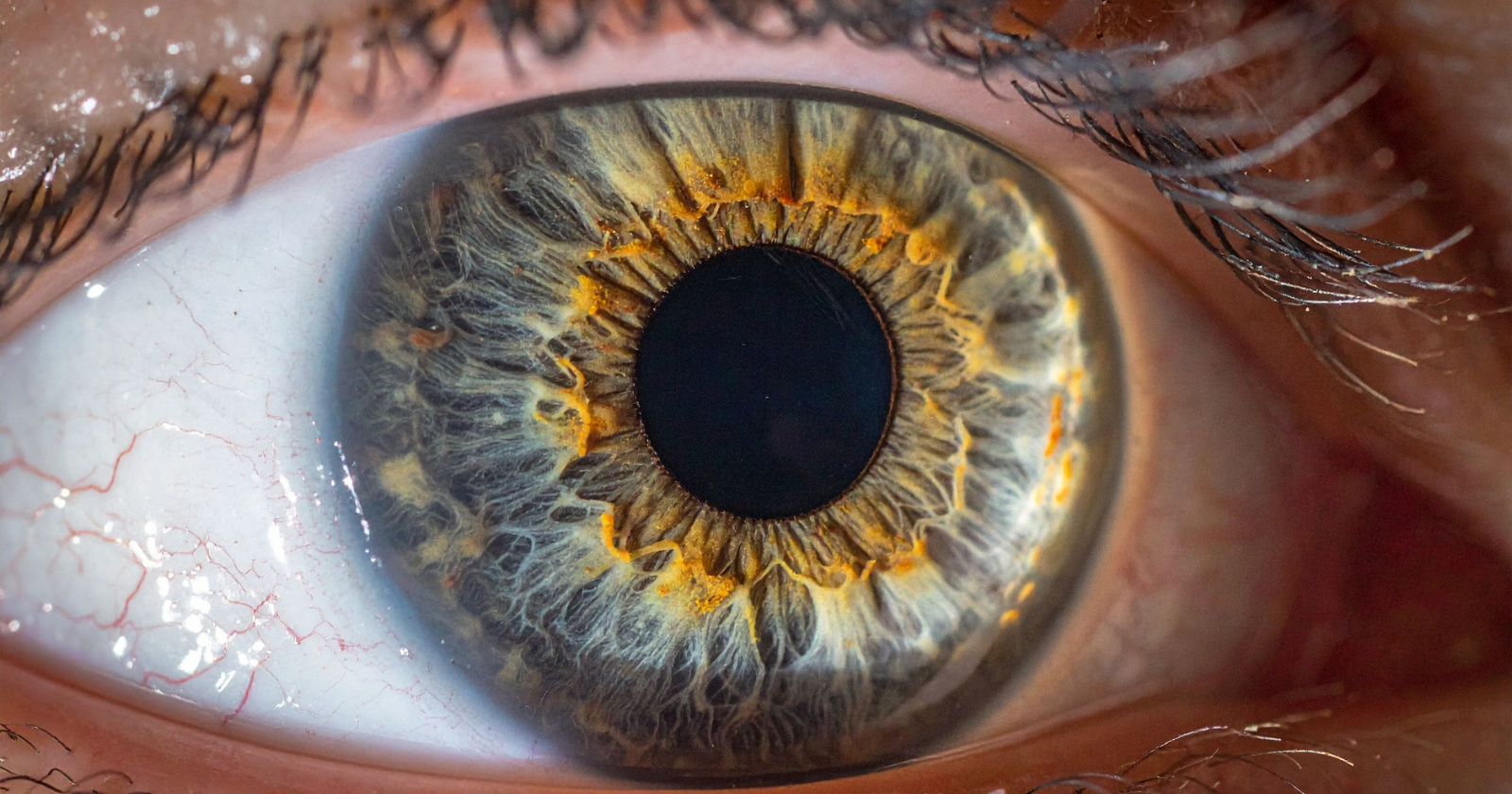
The eclipse of August 21, 2017 was, in the specific sense that interests astronomers, a normal eclipse. It was complete in a narrow strip of the continental United States, from Oregon to South Carolina, and partial in the rest of the country. However, public interest in him was extraordinary. Tens of millions of people bought or borrowed protective glasses. Millions more went out and looked at the sun anyway. Among the people out that afternoon was a 26-year-old woman from Staten Island, New York, named Nia Payne. She didn’t have protective glasses. What he did have was the same set of instincts that most human beings have when looking at the sun, which is that you can look at it for a fraction of a second without being hurt, and the moment it becomes uncomfortable you look away. Their city was within the partial coverage zone. About seventy percent of the solar disk was hidden behind the moon. She looked up for about six seconds. Then, uneasily, she borrowed what she thought was a pair of eclipse glasses from someone nearby. According to the subsequent evaluation of the ophthalmologists who would examine it, they were not the ISO-12312-2 certified filters that the American Astronomical Society had spent months warning viewers to use. Then he looked at the sun for another fifteen or twenty seconds through the inadequate glasses. When he entered, his left eye already had a blurry area in the center of his field of vision. Over the next two days, the blurred area took on a specific, unmistakable shape. A crescent, placed exactly where the moon had been when he looked at it, sitting in the middle of everything he was trying to look at. What was happening in the back of his eye According to the peer-reviewed case report published in JAMA Ophthalmology in December 2017 by Chris Y. Wu, Michael E. Jansen, Jorge Andrade, Toco YP Chui, Anna T. Do, Richard B. Rosen, and Avnish Deobhakta at New York Eye and Ear Infirmary at Mount Sinai and the Icahn School of Medicine at Mount Sinai, titled “Acute solar retinopathy photographed with adaptation optics, optical coherence tomography angiography, and en face optical coherence tomography,” Payne presented at Mount Sinai several days after the eclipse. His visual acuity was 20/20 in the right eye and 20/25 in the left, which by clinical standards was a relatively mild impairment. However, what his subjective experience described was much worse. In the center of his vision, the crescent-shaped scotoma sat over anything he tried to read, concentrate on, or recognize. What ophthalmologists were looking for and what they found was damage to the photoreceptor layer at the back of the retina. The retina is the thin layer of specialized neural tissue that lines the inside of the eyeball. Its outer layer is made up of photoreceptors, that is, individual cells that respond to light. The rods are responsible for peripheral and low-light vision. Cones handle color and fine detail, and are most densely concentrated in a small central region called the fovea, which is the specific patch of tissue you use every time you look directly at something. Solar retinopathy is what happens when photoreceptors are exposed to concentrated sunlight long enough to be damaged by two overlapping mechanisms. The first is a simple thermal injury. The lens of the eye focuses incoming light on the retina, and when the incoming light is sunlight, the focused spot is bright enough to raise the temperature of the tissue at that point above the level that the cells can tolerate. The second, and usually the most damaging, is a photochemical process. Ultraviolet light and short-wavelength visible light contain enough energy per photon to break specific molecular bonds within the light-sensitive proteins of photoreceptor cells, producing free radicals that damage surrounding structures even at temperatures below the thermal threshold. Photoreceptors, once destroyed, do not regenerate. The tissue does not grow back. To learn more about what happens to us during a solar eclipse, watch this video we made recently: The drawing and the image that corresponded to it. What the Mount Sinai team asked Payne to do, when he first arrived at their clinic, was to draw what he was seeing. Do not describe. Draw. On a sheet of paper, using an ordinary pen and pencil, he reproduced the shape of the blind area that was in the center of his vision. What he drew, in the contemporary record that survives, was a crescent. Specifically, a crescent oriented the same way the partial eclipse had been oriented when it faced the sun on August 21. The concave curve of the moon’s disk, imprinted in the visual field of a person who was no longer looking at the sun, looking at it from the center of everything he tried to look at. The next step, according to the primary source record of the case report, was to look inside his eye and see if the damage there matched the shape he had drawn. Doctors used a technique called scanning light ophthalmoscopy with adaptive optics, which is a high-resolution imaging method that corrects small imperfections in the shape of a person’s cornea and lens in real time, allowing the retina to be photographed with cellular resolution. The technology was originally developed for astronomy, to correct for atmospheric turbulence when photographing distant stars. Adapted for medical imaging, it allows doctors to see individual photoreceptor cells inside a living human eye. What the images revealed, in JAMA Ophthalmology figures accompanying the case report, was a region of destroyed photoreceptors in the central retina of Payne’s left eye and a milder version of the same pattern in his right. The destroyed region, in both eyes, was crescent-shaped. Its orientation matched, within the small tolerances of biological variation, the specific orientation of the drawing Payne had produced. The image matched the sketch and the sketch matched the eclipse. According to Mount Sinai Hospital’s own official statement on the case, published alongside the JAMA publication in December 2017, Dr. Avnish Deobhakta, lead author of the study, described the correspondence between the drawing and the images as unusually direct. What was visible in the adaptive optics scan was the specific imprint of a physical process that the eye’s own optics had performed on itself. The lens had focused the visible crescent of the sun on the specific area of the retina where the crescent had landed, and the light had destroyed the cells in that specific spot, and the pattern of destruction had appeared in Payne’s vision as the same crescent he had originally been looking at, only inverted, sitting between it and everything he was now trying to see. Payne’s condition, according to peer-reviewed literature covering subsequent years, has neither materially worsened nor improved. Solar retinopathy has no treatment. Photoreceptors do not regenerate. Over the years since the eclipse, he has retrained himself to use his right eye as dominant. Reading is still difficult. Screens require a lot of proximity. And in the center of his vision, on any shiny surface he looks at, the little pale crescent he drew for doctors at Mount Sinai in the fall of 2017 continues exactly where it always was, doing exactly what the case report predicted it would continue to do, silently, for the rest of its life. Kiran Athar is neither an ophthalmologist nor a medical researcher. He writes about science, medicine, and the ordinary corners of modern life where the two intersect, drawing on peer-reviewed research and primary source studies.