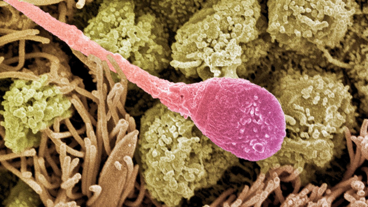
The load of a sperm includes more than the father’s DNA. RNA molecules that can contribute to early embryonic development also make the trip. Some of those molecules begin their journey outside of the sperm. Researchers have found that small protein fibers in semen can help RNA in the surrounding fluid enter sperm. In a new study published in Molecular Cell, the team showed that these fibers can bind to RNA, enter sperm, and help deliver that cargo to mouse embryos after fertilization. The fibers have an amyloid structure, best known for its association with Alzheimer’s disease. Structural biologist Anthony Fitzpatrick and physiologist William Mallen of Columbia University’s Zuckerman Institute, along with their colleagues, investigated how these fibers might help RNA enter sperm. “This is wild biology,” says Fitzpatrick. “Amyloids are most often thought of as a hallmark of Alzheimer’s, Parkinson’s, and other neurodegenerative diseases in the brain, but they also have functional roles throughout the body and continue to appear in unexpected places.” Amyloid describes the arrangement that proteins form when they are assembled into fibers. Those studied here are built from a fragment of a protein found in semen, different from the proteins that accumulate in Alzheimer’s. In semen, they form outside the cells of healthy men and can separate again. Some RNA molecules carry instructions for making proteins, while others help regulate gene activity, giving this cargo a potential role in early development. The researchers focused on a small fragment of an enzyme produced by the prostate, called prostatic acid phosphatase (PAP). The fragment carried a positive electrical charge, while the RNA carried a negative charge. When mixed in the lab, they stuck together and formed tiny droplets that turned into fibers containing RNA. On their own, the PAP fragments remained in solution. The addition of RNA brought them together and accelerated their assembly into fibers. That was the first surprise: the RNA helped assemble its own transporter. “The cargo is helping to build the vehicle that carries it,” Fitzpatrick told ScienceAlert. In human seminal fluid, the team also found small RNA molecules in a fraction containing amyloid fibers. Many identifiable sequences were fragments of transfer RNA, molecules normally involved in protein assembly. Finding RNA next to the fibers did not show whether the fibers could enter the sperm. Under some imaging conditions, a fiber attached to the surface of a cell can closely resemble one found inside it. Tiny gold particles attached to lab-made fibers allowed researchers to track them. They used bull sperm to obtain detailed images because the cells are relatively large. A technique that creates three-dimensional images of frozen cells, called cryo-electron tomography, revealed labeled fibers within the sperm. “Seeing those amyloid fibers inside the sperm was extraordinary to me,” Mallen says. “There were hints of this happening before in our other experiments, but actually seeing it completely stunned us.” Additional electron microscopy and live cell imaging supported the finding. Separate experiments showed that human and mouse sperm could also absorb the fibers. “The second surprise was seeing the sperm take those whole fibrils and move them deep into the cell, along the tail, through the midpiece and up to the head,” Fitzpatrick said. Amyloid fibrils (yellow) inside a sperm. (William Mallen/Fitzpatrick Laboratory/Columbia’s Zuckerman Institute) The journey continued in laboratory fertilization experiments using mouse sperm and eggs. Before fertilization, the team exposed the sperm to fibers made from fragments of human PAP. Those human protein fragments also appeared in the resulting two-cell mouse embryos. To track RNA loading, the researchers loaded the fibers with RNA containing heavier versions of carbon and nitrogen atoms. A technique that distinguishes molecules by mass allowed them to track these tags. More labeled RNA reached the embryos when fibers were present. The team tested whether the released RNA could work using RNA that carried instructions for a protein that emits green light. This protein served as a marker showing that the instructions had been used. The embryos were formed from sperm that were given RNA and whose fibers contained at least ten times more protein than embryos formed from sperm that were given the same RNA without fibers. Separate laboratory tests showed that a protein called clusterin accelerated the release of RNA from the fibers. Clusterin was also present in the embryos, although its role in the release of RNA within them has not yet been confirmed. The findings reveal a route by which RNA can travel from seminal fluid to sperm and then to the embryo. Understanding that pathway could also help researchers design new tools for delivering RNA. “There are metabolic disorders like insulin resistance that recent papers have shown can be transmitted through the paternal line in mice, and RNA is thought to be the way,” Mallen explains. “By learning how epigenetics-based disorders work, we can get closer to how to treat or prevent them.” The embryo delivery experiments used mice, lab-made fibers, and added RNA. Fitzpatrick said the team has not yet shown whether natural fibers, along with their usual load of RNA, change anything in an embryo. No human embryos or natural conceptions were tested, and uptake estimates varied depending on measurement methods. What happens to the fibers after they reach the embryo is still unclear. “We don’t know what happens to them in the embryo, whether they are removed, left in place or added even more,” Fitzpatrick said. The research has been published in Molecular Cell. This article was fact-checked by Rebecca Dyer and edited by Rebecca Dyer. While we take pride in our process, we are human. If you spot an error, please let us know.